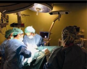
Hyperspectral imaging Cancer testing has now completed the prototype, which is expected to be tested in 2016

Hyperspectral Imaging Helps Identify Brain Tumors
Although malignant brain tumors in adults are only the thirteenth most common type of all cancers in the world , due to the difficulty in diagnosing individuals with the disease, they are the fifth most common cause of death from cancer in individuals under 65.
While it is feasible to effectively identify and remove tumors in other organs that have healthy tissue surrounding them, brain tumors infiltrate healthy tissue, making their edges indistinct and problematic for surgeons to identify. While techniques such as ultrasound and MRI (Magnetic Resonance Imaging) are deployed as aids to assist surgeons to differentiate between malignant and healthy tissue, no existing imaging system has been proved to be truly effective at doing so in the operating theater in real-time.
Hyperspectral imaging cancer detection
Now, however, a multidisciplinary European consortium composed of surgeons, pathologists, engineers and physicists is seeking to rectify that matter. Led by researchers at the Institute for Applied Microelectronics at the University of Las Palmas de Gran Canaria, the members of the consortium are readying a system called HELICoiD (HyperspectraL Imaging Cancer Detection) that will be able to precisely discriminate between healthy and malignant tissues in the brain in real-time. Having done so, the system may eventually enable surgeons to minimize the adverse effects of removing cells from the brain during surgical procedures.
Research and development activities are being carried out by four universities. Aside from the Universidad de Las Palmas de Gran Canaria, researchers at Imperial College London, the Universidad Politécnica de Madrid, and
Mines-ParisTech in France are also involved in the project. The consortium also includes Oncovision in Spain and Virtual Angle in the Netherlands, two SMEs with experience in medical imaging technology and software development, and Medtronic, a Spanish developer of surgical navigation systems.
ionizing system created by the European members of the HELICoiD consortium is able to discriminate between tissue types based on their chemical composition, a unique feature of the hyperspectral imaging technique. Although research on the project only started in 2014, a prototype system has been built and the core software that runs on the system has been written. The researchers are currently fine tuning the prototype and expect that a demonstrator will be ready for trials by the end of 2016.

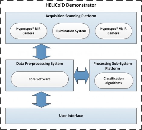
The HELICoiD system itself uses a pair of hyperspectral cameras from Headwall Photonics mounted on a platform situated above a patient that capture images of the brain during surgery. The area of interest is illuminated by a 150W Quartz Tungsten halogen light which produces broadband emissions in the 400 nm to 2200 nm spectral range. Due to the high temperature of the light, a fiber optic cable carries the light to a cold light emitter located in the platform with the cameras. As the brain is illuminated, one VNIR (visible and near-infrared) camera captures wavelengths in the 400-1000nm region of the spectrum, while the second NIR (near-infrared) camera captures wavelengths in the 900-1700nm range.
Datacube of images
Both hyperspectral cameras capture a large amount of data across a number of contiguous spectral bands over a wide spectral range. The result is what is known as a datacube: a stack of images collected at different wavelengths that contain both spatial and spectral information. To reduce the size of the data set without losing any valuable spectral and spatial information, and to ensure that the system will be able to operate in real time, the data from the cameras are transferred to an off the shelf processing unit for preprocessing prior to analysis.
Here, algorithms written by researchers in France, London and Las Palmas reduce the multi-band hyperspectral images to a set of smaller multi-spectral images with a limited number of bands. To do so, feature space reduction is performed on the data, whereby only those specific wavelength bands which contain data that are useful in discriminating between healthy and malignant tissue are retained by the system. In this way, algorithms can then quickly search for features in the reduced data set that are characteristic of cancerous tissue.
In order to identify the cancerous tissues in the brain, the HELICoiD researchers used a previously acquired dataset of hyperspectral images of healthy and cancerous tissue to train algorithms to detect differences in their spectral signature using machine learning techniques. When the HELICoiD system is used, the previously pre-processed images acquired during surgery are transferred to a high performance processing platform, such as an NVIDIA Tesla GPU (graphics processing unit), where the algorithms are highly parallelized to identify both cancerous and healthy tissue from the images in real-time.
Classifying and grading cancers
Having identified regions in the image that represent cancerous cells, the software will be able to classify the cancers into four different grades, depending on whether the cancer is benign, relatively slow growing, malignant, or highly malignant (tumor grades I, II, III and IV defined by the World Health Organization). It will also determine the areas in the brain where no cancer is present. The results from the analysis will be used by the visualization software running on the system to produce a pseudo-colored image that will highlight the differences between the cancerous and healthy regions in the image as well as the grade of any tumors that are found.
From the hyperspectral VNIR camera the system generates a RGB image of the brain during the surgical procedure. When the system is deployed in the theater, the pseudocolored image produced by processing the hyperspectral images will be overlaid with the RGB image. This will provide the neurosurgeon with an indication of how likely it is that specific areas of the brain are cancerous.
Prototype HELiCoiD systems are currently under test in the two hospitals involved in the project: the University Hospital of Southampton NHS Foundation Trust in the UK and the Hospital Doctor Negrín at Las Palmas in Spain. The results acquired from using the systems in actual hospital operating theaters during the following months will enable the researchers to refine the algorithms, control software and the user interface to ensure that the HELiCoiD works to its optimum capability.
Editor’s note: HELICoiD is a European collaborative project funded by the Research Executive Agency, through the Future and Emerging Technologies (FETOpen) programme, under the 7th Framework Programme of the European Union.
Written by Dave Wilson, Senior Editor, Novus Light Technologies Today
Labels: hyperspectral imaging,HELICoiD,brain tissue,imaging,Dave Wilson,FETOpen